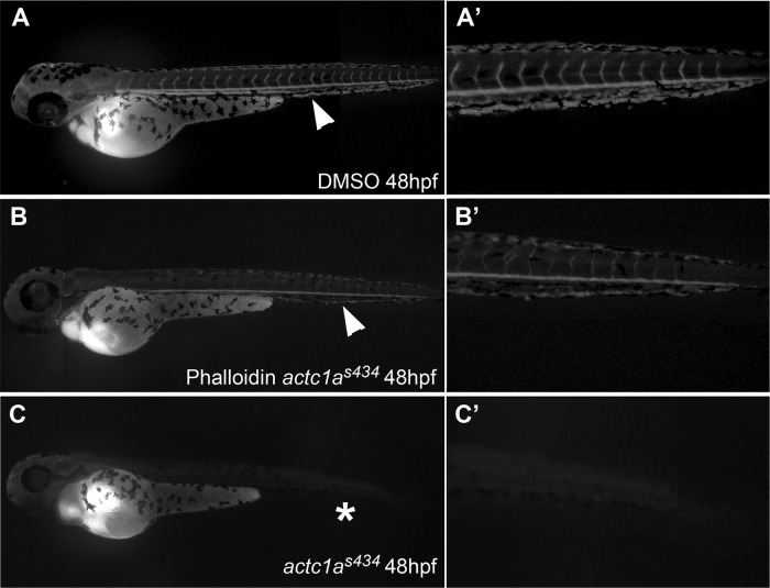
Fig 8.
Phalloidin treatment partially rescues circulation in actc1as434 mutants. Microangiography was performed at 48 hpf on wild-type sibling embryos (A), phalloidin-treated embryos that displayed pericardial edema, characteristic of s434 mutants (B), and untreated actc1as434−/− mutants (C). (A) In the wild-type embryos, injection of fluorescein-labeled dextran into the common cardinal vein (CCV) was circulated through the blood vessels of the body, as indicated by the arrowhead. Fluorescence is present in the dorsal aorta, the posterior cardinal vein, and intersegmental vessels (A′), shown in a magnification of panel A. (B) Phenotypically mutant embryos that displayed pericardial edema, alongside their wild-type siblings, were treated 24 to 48 hpf with a 2 μM dose of phalloidin oleate. Injection of dextran into treated mutant embryos showed a partial rescue in circulation, as the dye is present in the dorsal aorta (arrowhead). A magnified view of the trunk is shown in panel B′. (C) Injection of dextran into untreated mutant embryos demonstrated that these embryos do not have circulation, as magnification of their trunk shows there is no dye circulating in their vessels (C′). The images were taken using a 5× objective, with the anterior portion to the left and the dorsal side up.

