
Figure 4. Gene expression patterns are restored in treated embryos by quail neural crest cells.
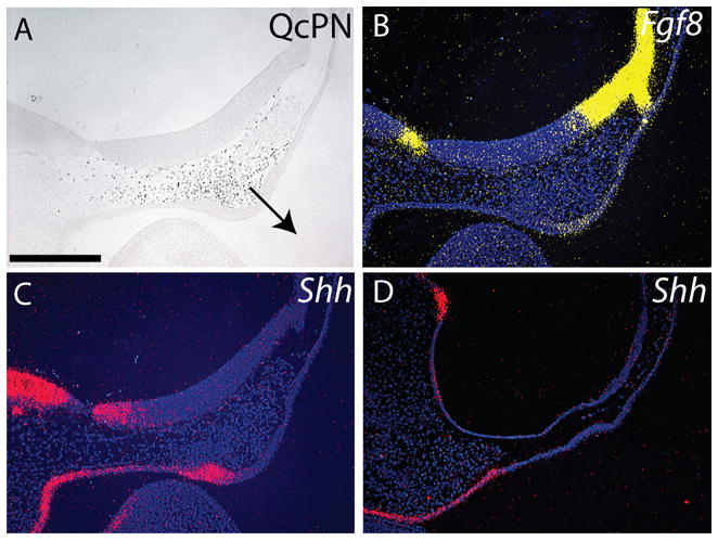
(A) Immunohistochemistry was used to assess quail mesenchyme at 24 hours after engraftment in chimeras. In this embryo, quail cells are located adjacent to the forebrain and are apposing the presumptive FEZ. The FNP exhibits signs of outgrowth (n=12, arrow=direction of growth). (B) Expression of Fgf8 in chimeric embryos (n=8). (C) The addition of quail cells to the FNP of treated embryos restored expression of Shh in the FEZ (n=8), (D) while embryos treated with SU5402 have no neural crest cells and no Shh expression in the ectoderm below the forebrain at HH22 (n=8). Scale bar: 250μm.