Figure 1.
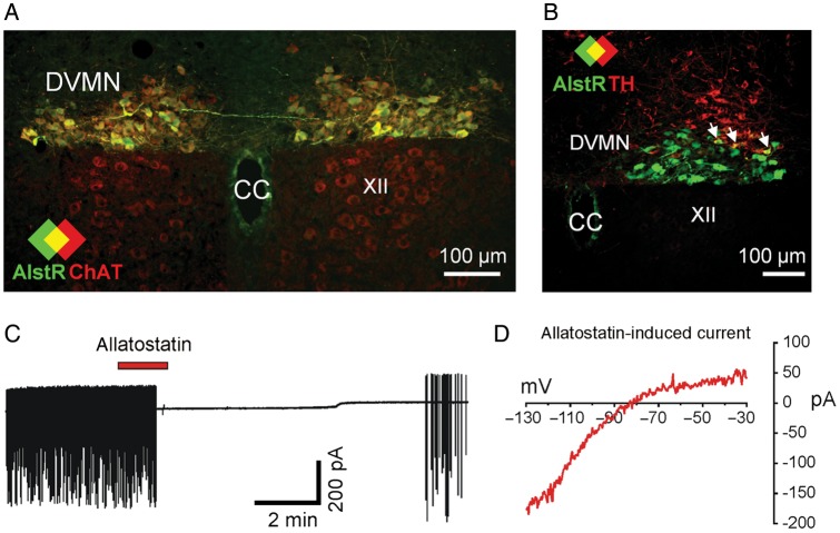
Genetic targeting and silencing of vagal pre-ganglionic neurones in the dorsal motor nucleus of the vagus nerve (DVMN). (A) Confocal image of the coronal section of the rat brainstem targeted to express allatostatin receptor (AlstR) in the DVMN. Figure illustrates a representative example of the distribution of choline acetyltransferase (ChAT)-positive (i.e. cholinergic) (red) DVMN neurones transduced to express AlstR/eGFP (green). Colocalization appears yellow. Bregma level −14 mm. XII, hypoglossal motor nucleus (ChAT-positive, but not expressing eGFP). CC, central canal; (B) a representative example of the distribution of AlstR/eGFP-transduced DVMN neurones in relation to the location of A2 noradrenergic cells identified by tyrosine hydroxylase (TH) immunohistochemistry (red). Along with strong expression of AlstR/eGFP in the DVMN only occasional noradrenergic neurones were found to be transduced. Colocalization appears yellow (arrows). Bregma level −13.8 mm. (C) Representative cell-attached recording from an AlstR/eGFP-positive DVMN neurone illustrating its rapid and reversible silencing in response to allatostatin (0.5 µM); (D) current–voltage relationship (IV) of allatostatin-induced current. Transduced DVMN neurone was voltage-clamped to −30 mV and hyperpolarizing voltage ramps to −130 mV (700 ms duration) were applied before and during allatostatin application. The displayed IV was obtained by subtracting the whole-cell IV obtained under control conditions, from that obtained in the presence of allatostatin.

