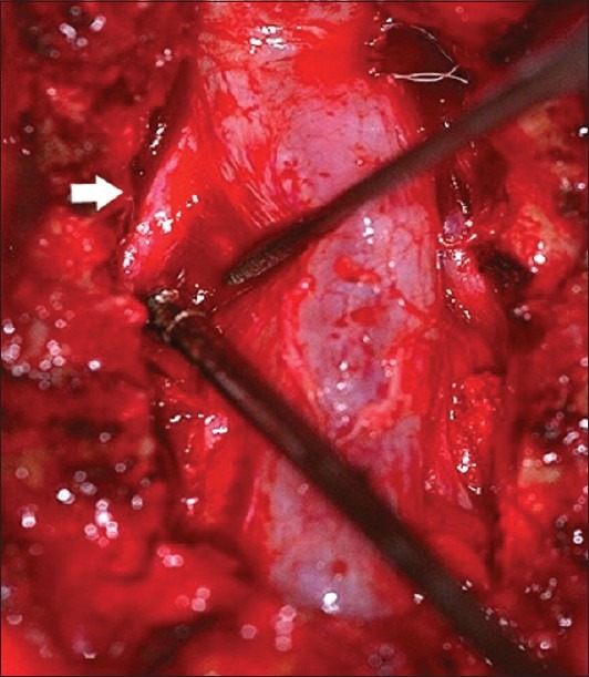
Figure 11.

Following excision of an L4–L5 massive left-sided synovial cyst extending to the L3–L4 level, this intraoperative photograph reveals the freed superiorly and foraminally exiting L4 nerve root (open arrow), and the decompressed thecal sac and inferior L5 nerve root. Additionally, the Penfield elevator and suction show the massive dimensions of the cavity previously occupied by the synovial cyst
