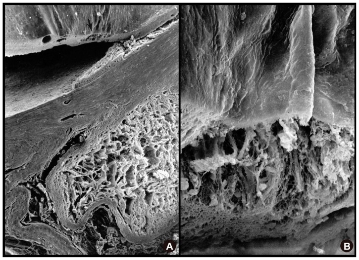
Figure 5.
SEM showing extensive cavernous atrophy of the optic nerve. (A) SEM showing extensive cavernous atrophy of the optic nerve with an intact subarachnoid space. (B) higher magnification SEM of cavernous atrophy of the optic nerve with a wall of intact glial tissue surrounding this area (upper portion of image).
Abbreviation: SEM, scanning electron microscope.

