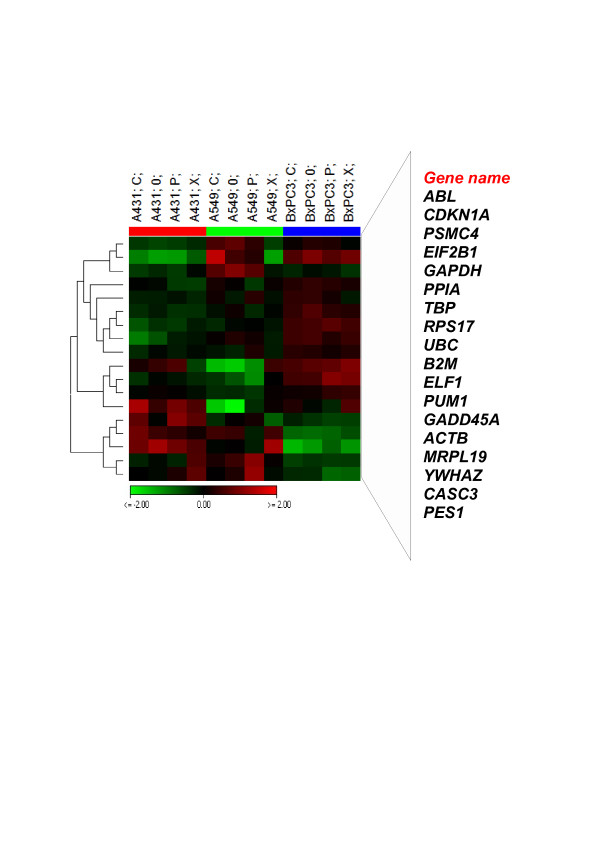
Figure 3.
Cell type specific regulation of ECGs. This Heat map represents 18 ECGs which are significantly regulated according to ANOVA between the three cell lines, p < 0.05. Relative expression of each gene were normalized to the average intensity of the gene over entire samples (virtual pool). Green denotes genes with relatively decreased expression while red denotes genes with relatively increased expression. Genes are hierarchically clustered by Pearson correlation coefficient using average linkage. Scale bar represent log2 expression level of ECGs. C = Carbon, P = Proton, X = Photon and 0 = Control. Expression levels of the ECGs in BxPC3 is different as compared to A431 and A549. A549 showed maximum variation among the samples.

