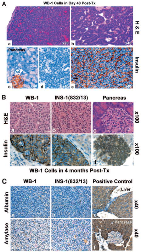
FIG. 5.
Histology and immunostaining of the explanted tissues. Paraffin sections of explanted tissues from 40 days (A) and 4 months (B) posttransplantation were stained with H&E (upper panels of A and B). The sections were immunostained with antibodies to insulin (1:500), amylase (1:200), and albumin (1:200) and incubated with matched secondary antibodies. The labeled proteins were visualized with DAB reagent. Lower panels of A represent insulin staining (c, pancreatic islet; d, negative control; e, insulin in the explanted tissue) in 40-day explanted WB-1 cells. Lower panels of B represent insulin staining on the explanted tissues containing WB-1 (left) cells, INS-1 (middle), and mouse pancreas tissue (right) as positive controls, as indicated in the figures. Each figure in B contains an internal negative control. C shows immunostaining of amylase and albumin in WB-1, INS-1, and positive controls (liver and pancreas), as indicated in the figure. Tx, transplanted.

