
FIG. 7.
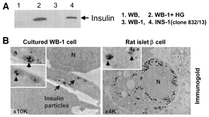
Detection of insulin and insulin secretory granules. A: Mature insulin was detected in the differentiated WB-1 cells (lane 2) by Western blot with anti-insulin antibody (1:500; Santa Cruz). All lanes were loaded with 50 μg protein except for INS-1 (loading only one-tenth protein [5 μg] to the well). B: Insulin secretory granules were detected by electron microscopy combined with immunogold labeling using anti-insulin antibody in an in vitro–differentiated WB-1 cell (B, left). Rat islet β-cells serve as the positive control (right). N represents the nucleus, and arrows indicate immunogold-labeled insulin particles.