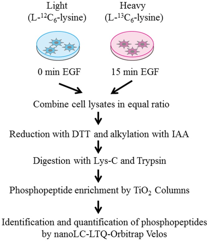
Figure 1. Schematic procedure for comprehensive identification and quantification of EGF-induced phosphoproteome based on SILAC technology.

Two populations of glioblastoma initiating cells were grown in media supplemented with normal (L-12C6-lysine) and stable isotopes (L-13C6-lysine), respectively. Each cell population is unstimulated or stimulated with 20 ng/ml EGF for 15 min, lysed, and combined in equal ratio. Phosphopeptides enriched through TiO2 columns are subjected to mass spectrometric analysis.
