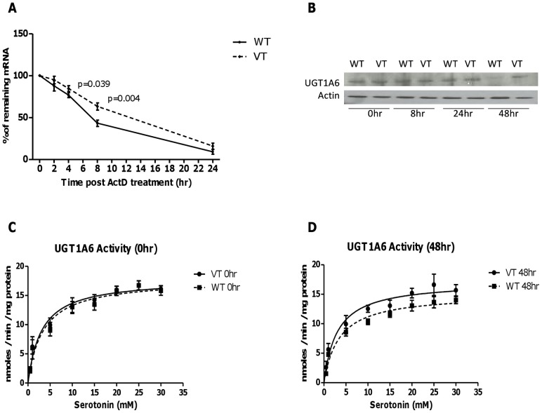
Figure 1. In vitro functional characterization of UGT1A6 105C>T polymorphism.
UGT1A6*1 and UGT1A6 105TT constructs were stably transfected into HEK293 cells. (A) Cells were harvested upon treatment with ActD at different time point up to 24 hours. The data plotted are time course changes in the remaining amount of UGT1A6 mRNA after ActD treatment. There was significantly higher mRNA level in UGT1A6 105TT than UGT1A6*1 transfected cells at 4 hours (p = 0.039) and 8 hours (p = 0.004) after treatment with ActD. (B) Western blot analysis. Cell lysate used was from 5×10⁵ cells at the indicated times (0, 8, 24 and 48 hours) after treatment with ActD. There was a down-regulation in protein expression in UGT1A6*1 as compared to UGT1A6 variant at 48 hours after treatment with ActD. UGT1A6 activity was assessed by evaluating the production of serotonin glucuronide using lysate from variant UGT1A6 (VT) and UGT1A6*1 (WT) at 0 hour (C) and 48 hours (D) after ActD treatment, with substrate concentrations varied from 0.5 to 30 mM serotonin. Activities are expressed as reaction velocity (nanomoles of serotonin glucuronide formed per minute per milligram of protein).

