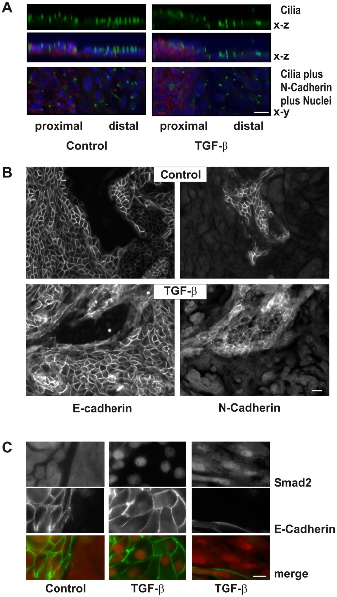
Figure 2. Polarization of primary epithelial cells.
(A) hPTECs were cultured in permeable filter inserts for 8 days and then further incubated with TGF-β for 7 days. Cilia were detected by staining with antibodies against acetylated tubulin. Merged images show an overlay of N-cadherin (red), DAPI (blue) and acetylated tubulin (green). X–y and x–z orientation is presented as indicated. Scale bar: 10 µm. (B) hPTECs were treated as described in A. Proximal and distal cells were distinguished by staining for N-cadherin and E-cadherin respectively. Scale bar: 20 µm. (C) Polarized hPTECs were treated with TGF-β for 2 h. Cells were stained with antibodies against E-cadherin and Smad 2/3. Cells not stained with E-cadherin were considered to be proximal hPTECs (right panel). Scale bar: 10 µm.

