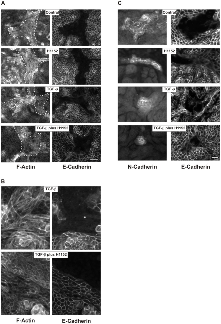
Figure 7. Structural alterations in polarized hPTECs.
(A) hPTECs were incubated in permeable filter inserts for 8 days to achieve polarization. They were further incubated with TGF-β (2 ng/ml) and H1152 (0.75 µM) for 7 days. Cells were stained for F-actin and E-cadherin. N-cadherin proximal tubular cells are visualized by F-actin only and are marked by the dotted lines (left panels). Scale bar: 50 µm. (B) Higher magnification of F-actin fibers in polarized cells treated with TGF-β or with TGF-β plus H1152 as described in 7A. Scale bar: 20 µm. (C) Cells were treated as in 7A. Proximal and distal cells were detected by antibodies directed against N-and E-cadherin, respectively. Scale bar: 50 µm.

