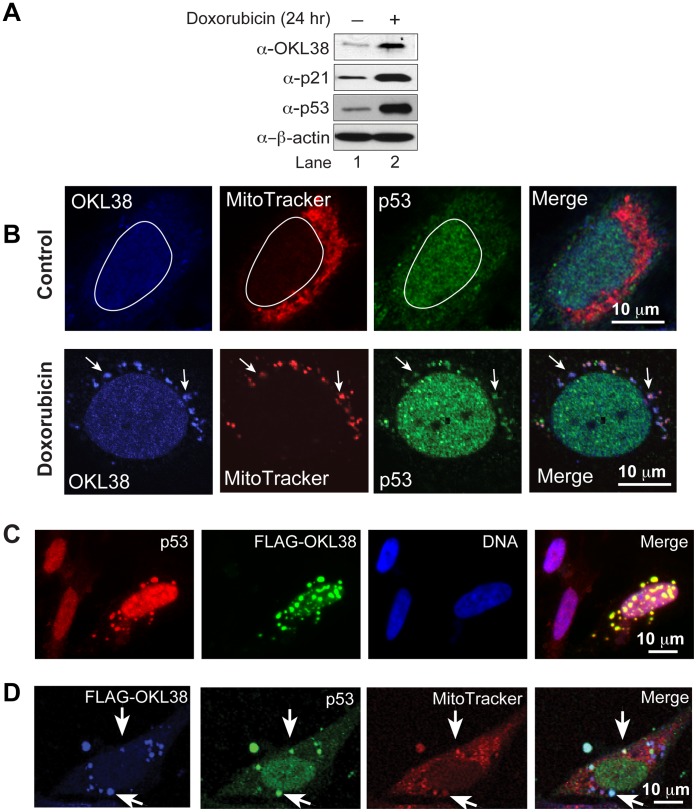
Figure 1. OKL38 and p53 colocalize in mitochondria.
(A) Changes in OKL38, p21, and p53 protein levels after doxorubicin treatment were analyzed by Western blot. β-actin was probed as a loading control. (B) Upper panels: before DNA damage, confocal images showed that OKL38 (pseudocolored blue) and p53 (green) were enriched in the nucleus outlined with a irregular white circle in U2OS cells. Lower panels: after DNA damage by doxorubicin, both OKL38 (blue) and p53 (green) were detected in mitochondria stained by the MitoTracker Red dye. Arrows denote the mitochondria spots with OKL38 and p53 staining. (C) Fluorescent microscopy images showed that p53 (red) and OKL38 (green) colocalized in large speckles outside of nucleus (DNA staining in blue). (D) Confocal images showed that FLAG-OKL38 (blue) and p53 (green) colocalized with mitochondria (red) after FLAG-OKL38 overexpression in U2OS cells. Arrows denote the large mitochondria speckles with both FLAG-OKL38 and p53 localization.

