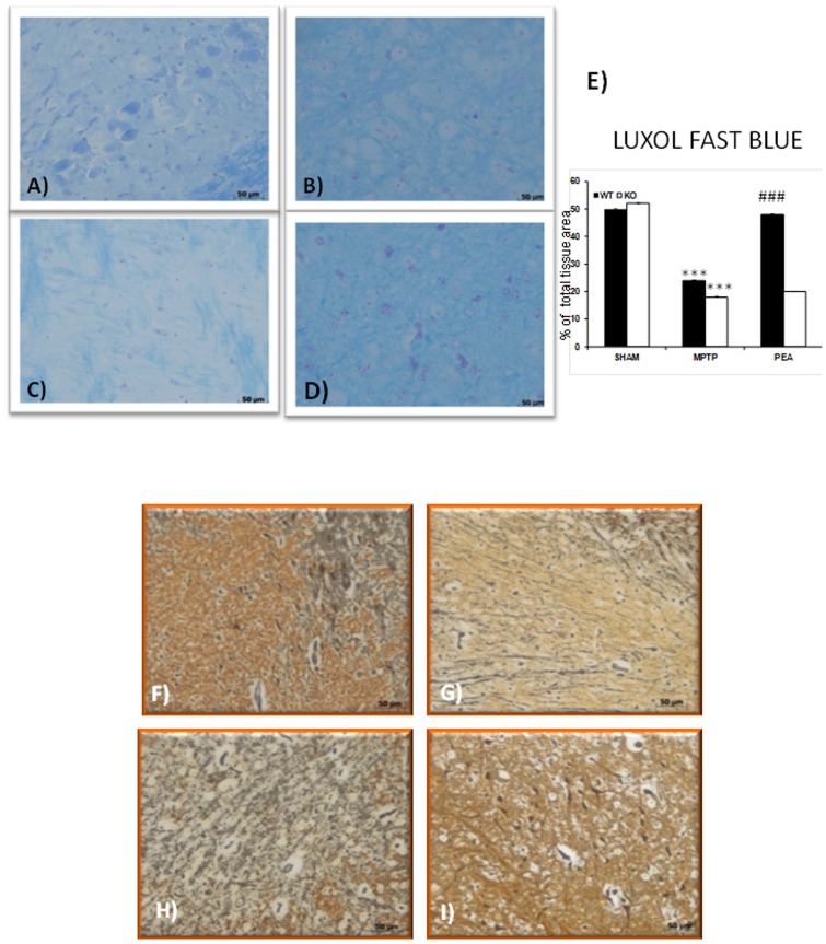
Figure 9. Effect of PEA on myelin structure and presence of granule-laden astrocytes (Gomori stains).
In sham PPARαKO animals (A and E), myelin structure was clearly stained by Luxol fast. At 8 days after MPTP injection in PPARαWT mice (B and E), a significant loss of myelin was observed. Myelin degradation was further progressed in PPARαKO mice and myelin negative area was defined in SN (C and E). The treatment with PEA significantly restored in PPARαWT the myelin presence (D and E). In addition, in sham PPARαWT mice (data not shown) or sham PPARαKO mice (F) a significant presence of astrocytes exhibiting an affinity for chrome-alum hematoxylin and aldehyde fuchsin was observed in the SN as well as in the vessels. On the contrary, a significant alteration of the Gomori positive localization was observed in the SN from MPTP-injected PPARαWT mice (G). A more alteration of Gomori positive localization was observed in MPTP injected-PPARαKO mice (H). PEA treatment reduced the alteration of the Gomori positive localization in PPARαWT mice (I).

