
Figure 4. Infarct volume was reduced by AFS cell transplantation.
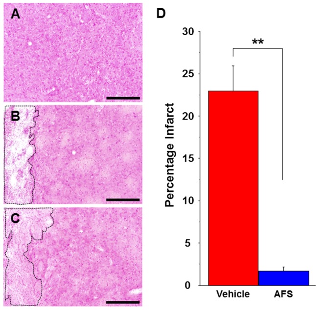
Stronger H&E staining is found in the intact striatum (A) compared to the vehicle-infused stroke animals (B) and AFS cell-transplanted stroke animals (C). Infarct volume is significanly reduced in the AFS cell-transplated stroke animals (C) compared to vehicle-infused stroke animals (B). The striatum in the AFS cell-transplated stroke animals is clearly preserved compared to that of vehicle-infused stroke animals (A-D). Quantitative analyses revealed that percentages of the infarct volumes of rats receiving AFS cell transplants are significantly reduced (**p<0.01) (D). Data are shown as percentages of the infarct volumes present in the ipsilateral hemisphere relative to the contralateral hemisphere. Bars represent the mean ± SEM. Scale bars = 200 µm. Black dotted box represents the infarct area.