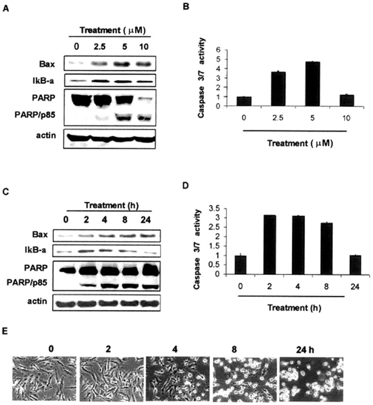
Figure 3. Dosage and kinetic effects of WA on apoptosis induction in human MPM cells.
H2373 cells were treated with different concentrations of WA for 16 h (A, B), or 10 µM WA for various times (C–E), followed by Western blotting analysis for levels of Bax, IκB-α, PARP, and actin proteins as detailed in methods (A and C). B, D, Caspase 3/7 activation was determined from by WA treated cell lysates as in methods. Bars, SD. E, Photomicrographs showing apoptosis-associated morphological changes in WA-treated MPM cells for indicated times. 0, Control, DMSO treatment.

