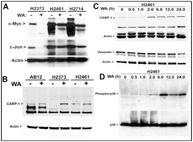
Figure 4. WA suppresses MPM survival and metastasis promoting genes while enhances expression/activation of pro-apoptotic genes.
MPM cells were either untreated (DMSO; denoted as −) or treated with 10 µM WA (denoted as +) and cell lysates were analyzed by western blotting as in methods. Levels of c-myc, c-Jun (A), and CARP-1 (B) proteins were determined in the cells treated with WA for 12 h. In addition, MPM cells were either untreated (DMSO, denoted as 0) or treated with 10 µM WA for the indicated time periods, and the levels of CARP-1, vimentin (C), and phospho-p38 (D) were determined by western blotting. The membranes in panels A–C were subsequently analyzed for levels of actin to assess protein loading, while the membrane in panel D was probed for expression of total p38.

