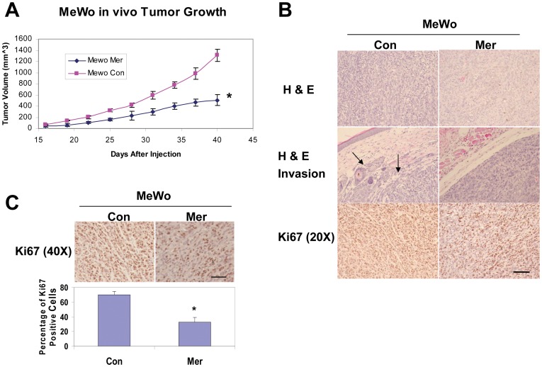
Figure 5. Increased merlin levels inhibit in vivo melanoma growth and invasion.
A, Subcutaneous growth rates of the melanomas derived from 1×10⁶ transduced MeWo cells as indicated. Numbers are the mean tumor volumes (mm3) +/− SD. * = p-value <0.05, n = 6. B, H&E and IHC staining with an anti-Ki67 antibody of the tumor sections derived from MeWo Mer or MeWo Con cells. Bar, 200 µm. C, Quantification of the percentage of Ki67 positive cells in the tumor sections derived from MeWo Mer and MeWo Con cells. Bars represent the means of 15 randomly selected 40X microscopic fields. *denotes a p-value <0.05. Bar, 200 µm.

