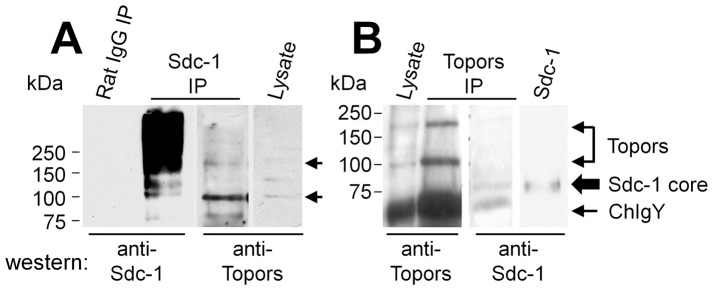
Figure 4. Co-immune precipitation of Sdc-1 and Topors from lysates of NMuMG cells.
(A) Total cell lysates prepared from NMuMG cells were immune precipitated (IP) with non-immune rat IgG (lane 1) or rat anti-Sdc-1 antibody (lane 2 and 3) and then western blotted for Sdc-1 (lanes 1–2), and after stripping, re-probed with anti-Topors (lanes 3). Total cell lysate (lane 4) was included as a control for anti-Topors immunoreactivity. Arrows mark major co-precipitated Topors bands (110 and 175 kDa). (B) Non-immune chicken IgY-preadsorbed total cell lysates of NMuMG cells were immunoprecipitated with chicken anti-Topors, and immune precipitates were heparinase- and chondroitin ABC lyase-digested prior to SDS-PAGE and western blotting. Lysate (lane 1) and Topors immunoprecipitates (lane 2 and 3) that were probed on western blots for Topors (lanes 1 and 2) had Topors bands (110 and 175 kDa bands, double arrow), or for Sdc-1 (lane 3), which gave a band of ∼85 kDa (large arrow, lane 3 and 4), similar in size to a band present in a Sdc-1-positive control (lane 4), prepared by ion-exchange chromatography from the medium of NMuMG cultures. Lower MW heavy bands represent chicken IgY (NChIgY) from the immune precipitate (arrow). Lanes 1–3 are from the same blot with lane 3 representing a stripped and re-probed lane of the Topors immune precipitate run in parallel to that shown in lane 2. Lane 4 represents an adjacent lane from the same blot, at a longer exposure time.

