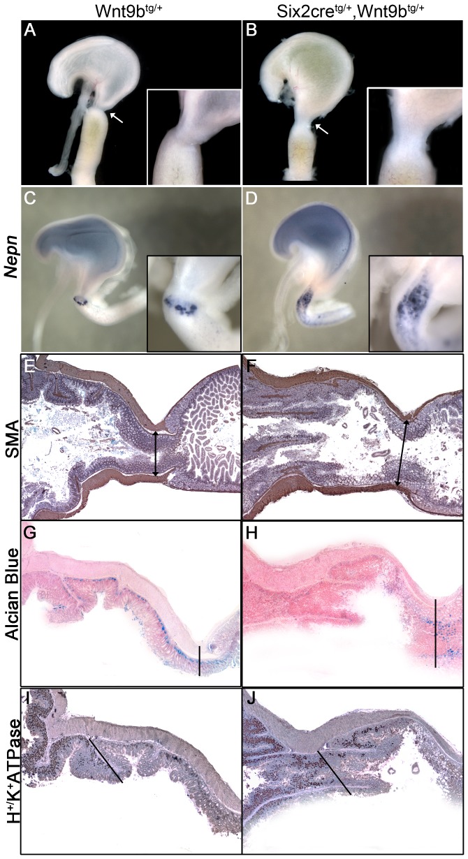
Figure 6. Duodenogastric reflux and pyloric sphincter abnormalities in Six2-cretg/+, Wnt9b tg/+ double heterozygotes.
(A, B) Yellow amniotic fluid refluxed into the stomach of E18.5 double heterozygotes, but was properly retained in the intestine in control animals. A lack of constriction (arrow) and an increased diameter (inset) suggests that the reflux is due to a malformed pyloric sphincter. (C, D) Whole mount staining for Nephrocan mRNA delineates a thin band demarking the sphincter at E16.5 that is expanded in the double heterozygotes. (E, F) Smooth muscle actin staining in sections from control adult stomach demonstrates a thick muscular layer that leads to the sphincter. Double heterozygotes exhibit a thickened muscular layer; however it does not coalesce at the point of highest constriction. Vertical lines mark the transition from antral stomach (left side) to duodenum (right side) in E–H. (G, H) Alcian blue stains mucosal cells in the antral stomach in control sections and these cells are absent in the double heterozygotes. Mucosal cells in the intestine are stained normally. (I, J) Staining for the H+/K+ ATPase detects parietal cells in the forestomach that are excluded from the distal stomach. These cells are found throughout the distal stomach and pylorus in double heterozygotes. Sections depicted in panels I and J highlight the transition between forestomach and distal stomach (diagonal line) and do not include the duodenum.

