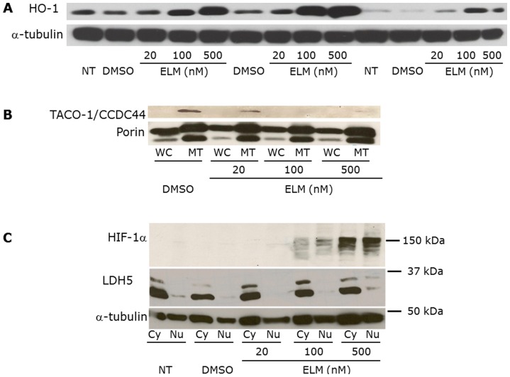
Figure 2. HO-1, TACO-1, and HIF-1α expression in Elesclomol-treated melanoma cells.
(A) Whole-cell (WC), (B) mitochondrial and WC, and (C) nuclear (Nu) and cytoplasmic (Cy) lysates, prepared from WM1158 metastatic melanoma cells following treatment with increasing doses of Elesclomol (ELM). Controls were WM1158 melanoma cells that received the drug vehicle DMSO, or no treatment (no tx). The blots were probed with antibody to HO-1, TACO-1, HIF-1α, or α-tubulin, which served as loading control. LDH5 was used as a cytoplasmic protein control.

