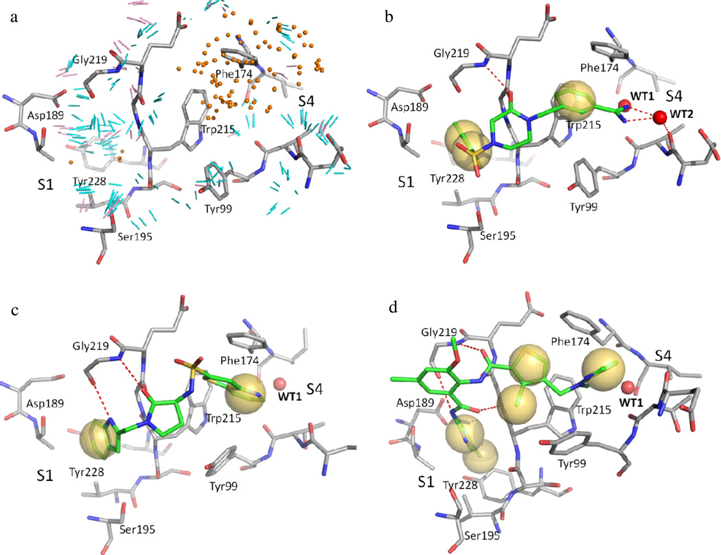
Figure 2.
Protein pharmacophores identified in the active site of fXa:1NFU (a) compared with the interaction features between fXa and its co-crystallized ligands (b: RRP in 1NFU, c: PR2 in 1F0S, d: XLD in 1MQ6). The coordinates for binding site residues shown as grey sticks represent the minimized crystal structure of fXa. a: The pharmacophore elements representing potential ligand functional groups are coded as follows: orange dots as hydrophobic groups, cyan lines as hydrogen bond donor groups, pink lines as hydrogen bond acceptor groups. b–d: Red dash, hydrogen bond interaction; Yellow sphere, hydrophobic interaction. A conserved water molecule (WT1) observed in all three crystal structures and a mediating water molecule in 1NFU (WT2) are shown as red spheres.

