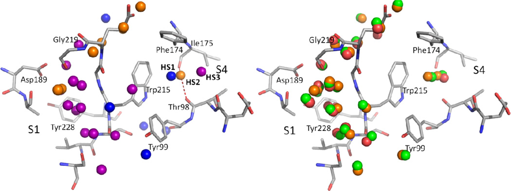
Figure 5.
Hydration sites identified in the binding site of factor Xa. For clarity, hydration sites that are not accessible to the ligands or on the protein surface are removed. The protein structure shown is the minimized crystal structure of 1NFU. In the left panel, only hydration sites identified from 1NFU are shown. Hydration sites that contribute both entropically and enthalpically when replaced by ligands are shown in blue. The hydration sites whose entropic gain surpasses the enthalpic loss when replaced by ligands are shown in purple. The hydration sites whose enthalpic loss is larger than the entropic gain when replaced by ligands are shown in orange. In the right panel, the hydration sites identified from three different starting structures of factor Xa were overlaid (red: 1F0S, green: 1MQ6, orange: 1NFU).

