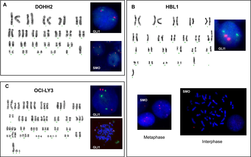
Figure 4. G-banding karyotype and FISH assays for DOHH2, HBL1 and OCI-LY3 cells.
In DOHH2 cells, G-banding showed trisomy 7 and 2 chromosomes 12. Dual-color interphase FISH confirmed the presence of 3 copies of SMO (ratio 1:1 with the centromere probe) and 2 copies of GLI1 in this cell line. In HBL1 cells, G-banding showed one intact chromosome 7 and a der(12) having the other chromosome 7. The presence of 2 copies of SMO gene was confirmed by interphase and metaphase FISH assays. In OCI-Ly3, G-banding show trisomy 7 and 12 and dual-color interphase and metaphase FISH studies confirmed that the presence of extra copies of SMO and GLI1 genes were associated with chromosomal aneuploidies (ratio 1:1 with centromere probes).

