Figure 7.
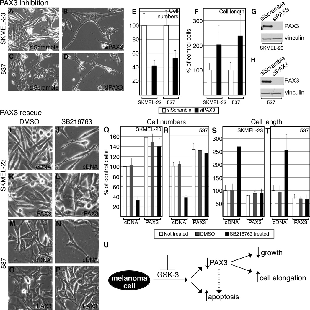
PAX3 knockdown replicates cell phenotypes of GSK-3 inhibition (A–H) and PAX3 over-expression rescues these phenotypes (I–T). A–D, overall morphology of SKMEL-23 (A–B) and 537 (C–D) transfected with siScramble or siPAX3. Cells transfected with siPAX3 exhibited cell number reduction compared to siScramble at 72 hours. E, quantification of cell number reduction with siPAX3. Cells transfected with siPAX3 were counted 72 hours post-transfection and compared to siScramble (set at 100%). Values are means +/− s.d. (n=600 cells). F, quantification of cell length upon transfection with siScramble or siPAX3. 10 cells were measured from 6 groups. The siScramble was expressed as a percentage of the control group (average set at 100%). Values are means +/− s.d. (n=60 cells). G–H, siPAX3 reduces PAX3 levels in total cell lysate of SKMEL-23 (G) and 537 (H) compared to siScramble. Blots were probed with PAX3 antibody and vinculin as a loading control. I–P, SKMEL-23 (I–L) and 537 (M–P) cells were transfected with pcDNA3 (I–J, M–N) or pcDNA3-PAX3-HA (K–L, O–P) and treated with DMSO (I,K,M,O) or SB216763 (J,L,N,P) for 72 hours and observed for cell population and overall morphology. Q–R, quantification of cell numbers in I–P. SKMEL-23 and 537 cells transfected with either pcDNA3 or pcDNA3-PAX3-HA were left untreated, treated with DMSO, or with SB216763 for 72 hours and counted and compared to the non-treated control (set at 100%). Values are means +/− s.d. (n=600). S–T, quantification of cell length from I–P. For each treatment, 10 cells were measured from 6 groups. The DMSO- and SB216763-treated groups were expressed as a percentage of the untreated cells (average set at 100%). Values are means +/− s.d. (n=60). U, summary schematic of the response of melanoma cells to GSK-3 inhibition. A loss of GSK-3 activity resulted in an overall reduction of PAX3 levels, a decrease in cell growth and survival, and cellular morphology changes in some but not all of the cell lines tested. While the effects on cell growth and elongation are linked to a loss of PAX3 in this study, this correlation could not be established between PAX3 loss and apoptotic induction. PAX3-dependent apoptosis has been reported in melanoma cells, however, and is indicated with a dotted arrow (20, 24).

