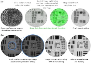
Fig. 2.
Snapshot spectral encoding calibration procedure. (a) A datacube of a high resolution 1951 USAF target is acquired. The fiber bundle pattern is removed from each spectral image using interpolation based on fiber core centers. Spectral images are then registered. All spectral images are combined by taking the maximum fiber core intensity at each image coordinate. Effectively, void spaces between fibers in one spectral image are filled with fiber core intensity from other spectral images. Any remaining void spaces are filled with interpolation. (b) An image from a traditional fiber bundle endomicroscope5,6 has a pixelation effect. The result of snapshot spectral encoding shows improved resolution and contrast, comparable to an image obtained directly from a microscope stage. The dotted circle represents the region of interest shown in (a).

