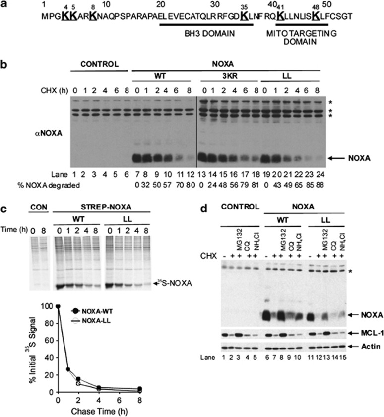
Figure 1.
NOXA is degraded by proteasomes in a lysine-independent manner. (a) Primary sequence of human NOXA. Lysine residues are shown as large bold text. BH3 and mitochondrial-targeting domains are underlined. (b) NOXA is degraded via a lysine-independent pathway. Cells were transfected as indicated, and NOXA degradation assessed by immunoblotting and quantified using Odyssey Image analysis (LI-COR, Cambridge, UK). (c) NOXA-WT and -LL have similar basal turnover rates. Cells transiently expressing Strep-tagged NOXA-WT or -LL were labeled with a 35S-Cys/Met mixture for 1 h, and chased with unlabeled Cys and Met for the indicated times. Strep-tagged proteins were captured on streptactin beads. Gels were subjected to autoradiography and 35S-NOXA quantified using a phosphoimager. Results shown are from one experiment typical of two. (d) Degradation of NOXA-WT and -LL is proteasomal-dependent. Cells were transfected for 48 h and treated for the final 12 h with DMSO, MG132 (25 μM), chloroquine (200 μM) or NH4Cl (20 mM), and NOXA degradation assessed. Asterisks denotes non-specific bands

