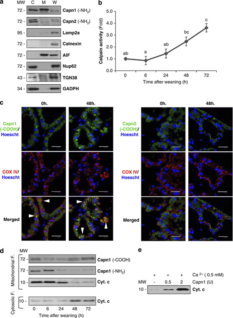
Figure 3.
Mitochondrial effects of calpain activation. (a) Immunoblot of the cytoplasmic (C), mitochondrial (M) and whole tissue (W) extracts with antibodies against calpain-1 and -2 in control lactating glands. To assess the purity of mitochondrial fractions, western blot analysis with antibodies against other organelle markers was performed: Lamp2a (lysosomal marker), Calnexin (endoplasmic reticulum marker), AIF (mitochondrial marker), TGN38 (Golgi apparatus), nucleoporin p62 (nuclei) and GAPDH (cytosolic marker). (b) Determination of calpain activity in mitochondrial extracts from control and weaned glands at different times of involution. All values are shown as means±S.E.M. ANOVA was performed for the statistical analysis, and different superscript letters indicate significant differences, P<0.05; the letter ‘a' always represents the lowest value within the group. (c) Mammary tissue sections from control lactating (0 h) or 48-h-weaned mice were fixed and immunostained with anti-COX IV (red) and calpain-1 or -2 (green) antibodies. Nuclei were visualized by Hoeschst-33342 (blue). Scale bar: 20 μm. (d) Western blot analysis of mitochondrial or cytosolic fractions at different times of weaning (0, 6, 24, 48 and 72 h), showing CAPN1 cleavage of the amino-terminal residues and subsequent cytochrome c release from the mitochondria to the cytosol. (e) In vitro treatment of isolated mitochondria from lactating mice with different units of recombinant calpain-1 in the presence of calcium. After 15-min incubation at 37 °C, mitochondria were pelleted and the amounts of cytochrome c released to the supernatant were assessed by western blot

