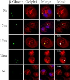
Fig. 5.
Glucans are trafficked to the Golgi apparatus in murine macrophages. Thioglycollate-elicited macrophages were incubated with fluorescent-labeled β-glucan for 3 h at 4°C, and then for 0 to 24 h at 37°C. The cells were stained for Golgi by using Golph4 antibody, and the images were obtained by confocal microscopy. The numbers shown in white in the masked images (Mask column) are the mask intensity rate for the images and indicate the degree of colocalization. A mask intensity rate of ≥40% was considered indicative of colocalization. Representative maximum projection images of four to eight replicates from two to four independent experiments are shown.

