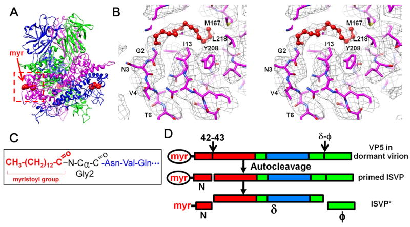
Figure 5. Myristoyl Group of the Primed VP5 in the ISVP.

(A) Positions of three myristoyl groups (red balls) near the base of the VP5 trimer. See also Movie S4.
(B) Cryo-EM density (gray mesh) of the base region in Figure 5A (red box), superimposed with its atomic model (magenta), showing the myristoyl group (red balls and sticks) inside a hydrophobic pocket. See also Movie S4.
(C) Amide linkage of the myristoyl group to Gly2.
(D) Changes of the penetration protein VP5 from a stable, dormant state to a metastable, primed ISVP state and subsequently to the ISVP* state. Red, base domain (2-242); green, linker domain (243-286 & 485-648) and cyan, jelly-roll domain (287-484). The myristoyl group (myr) in the hydrophobic pocket is enclosed by an ellipse; the released myristoyl group has no ellipse.
