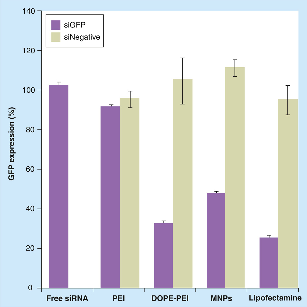
Figure 2. Effect of polyethylene glycol/lipid layer in micelle-like nanoparticles silencing efficacy.
c166-GFP cells (stably expressing GFP) were treated with the formulations prepared with GFP-targeted siRNA or nontargeted siRNA at PEI nitrogen/nucleic acid phosphate ratio of 16. The siRNA concentration was 100 nM. After 4 h of incubation, complexes were removed and cells were incubated for 48 h. Cells were trypsinized and analyzed by flow cytometry. The downregulation of GFP was measured by the decrease in the mean fluorescence of the treated cells compared with nontreated and expressed as a percentage of nontreated control cells. Data are expressed as the mean ± standard deviation (n = 3).
DOPE: Dioleoylphosphatidylethanolamine; GFP: Green fluorescent protein; MNP: Micelle-like nanoparticle; PEI: Polyethylenimine.

