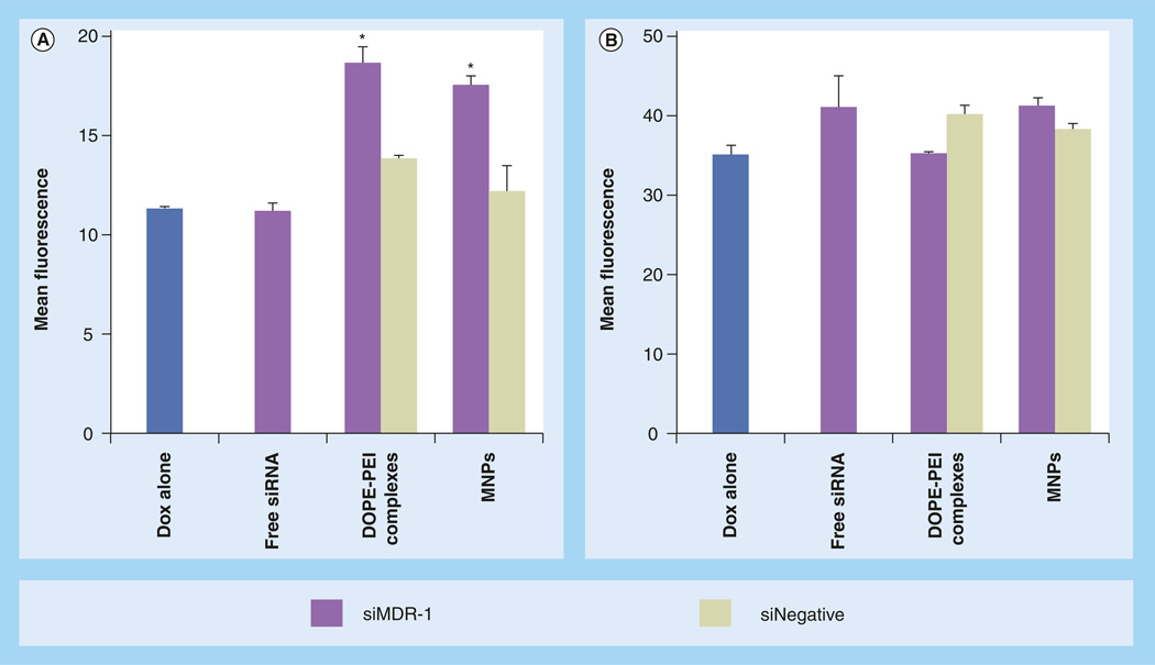
Figure 4. Intracellular doxorubicin levels.
In (A) resistant and (B) sensitive MCF-7 cells mediated by DOPE-PEI complexes and MNPs. MCF-7 resistant and sensitive cells were treated with formulations prepared either with siRNA targeting MDR-1 or scramble siRNA. Control cells were treated only with medium. After 4 h, medium was exchanged. Cells were reincubated for 48 h. Cells were then treated for 1 h with doxorubicin (5 µg/ml), then washed, trypsinized and analyzed by flow cytometry. The accumulation of doxorubicin within the cells was measured by the increase in the mean fluorescence. Note ordinate scale differences. Data are expressed as the mean ± standard deviation (n = 3).
*p < 0.001 versus Doxalone.
DOPE: Dioleoylphosphatidylethanolamine; Dox: Doxorubicin; MNP: Micelle-like nanoparticle; PEI: Polyethylenimine; siNegative: Scrambled siRNA; siMDR: siRNA targeting MDR.

