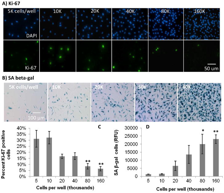
Figure 2.
Effect of increasing cell confluence on macrophage proliferation in culture. Fluorescence and color micrograph images of: A) DAPI and Ki-67, and B) SA beta-gal staining, respectively, in primary BMMΦs with increasing confluence on tissue culture plastic. Graphical representation of C) percent positive cells for Ki-67, and D) relative fluorescent signal, from lysed fluorescent SA beta-gal assay, showing increased SA beta-gal with increasing confluence in BMMΦ cultures. These data show decreased percent proliferative cells as seen by Ki-67 and an increase in SA beta-gal with increasing cell density. These data also confirm the ability of SA beta-gal to represent non-proliferative cells and the decrease in proliferative capacity of macrophages with increasing concentration. One-way ANOVA with post-hoc Dunnett Multiple Comparisons Test was applied, comparing all concentrations the 5K condition. Significance is noted as p<0.05 * and p<0.001 **.

