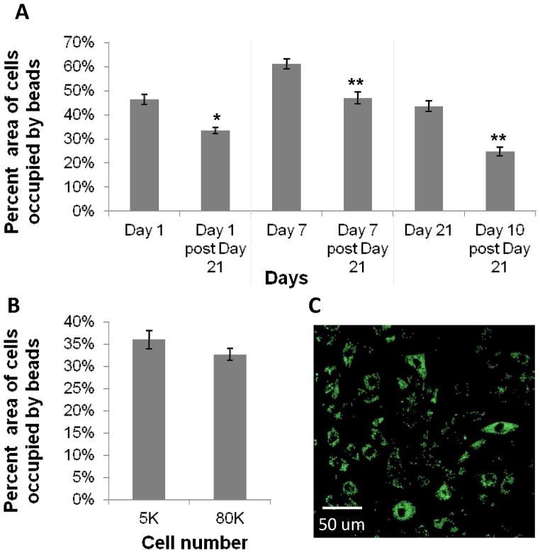
Figure 4.
A) Bead phagocytic uptake in macrophages passaged after 21 days (shown in Figure 3 to be senescent) and cultured for 10 days were compared with bead uptake of their corresponding time point prior to 21 days. Data show a reduction in phagocytosis in cells at time points past 21 days. B) Bead uptake in macrophages at low and high confluency, indicating no significant change in phagocytosis at high confluence (shown in Figure 3 to be quiescent). One-way ANOVA with post-hoc Student’s t-test was applied, comparing post passages to the prepassaged condition (i.e. Day 1, 7, and 21). Significance is noted as p<.05 * and p<.001 **. C) Representative confocal image of BMMΦs with internalized beads. This image is an overlay of 10 z-sections through the cell, indicating beads are internalized by phagocytosis.

