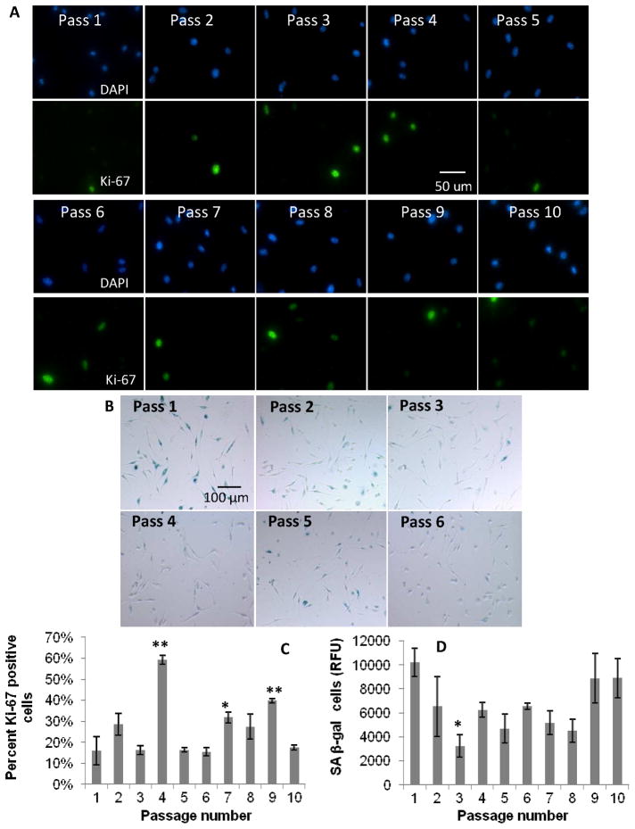
Figure 6.
Effect of serial passaging on BMMΦ proliferation. Fluorescence and color images of: A) DAPI and Ki-67, and B) SA beta-gal staining, respectively, in primary BMMΦ cultures with increasing passage numbers. Graphical representation of: C) percent positive cells for Ki-67 markers, and D) SA beta-gal (from fluorescent senescence assay) with increasing passaging in BMMΦs. These data show no dramatic increasing or decreasing trends in senescence with increased passaging. One-way ANOVA with post-hoc Dunnett Multiple Comparisons Test was applied, comparing all passages to the passage 1 condition. Significance is noted as p<0.05 * and p<0.001 **.

