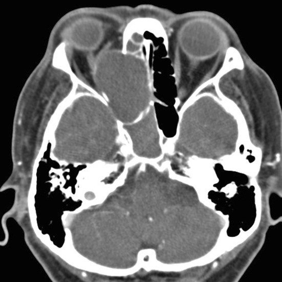
Fig. 1.

Axial images of CT scan of orbits with contrast demonstrating a 4.4 × 4.2 × 3.3 cm mass consistent with a right ethmoid mucocele. The medial orbital wall has expanded and mass effect on the medial rectus and optic nerve is noted. Right-sided proptosis is also present
