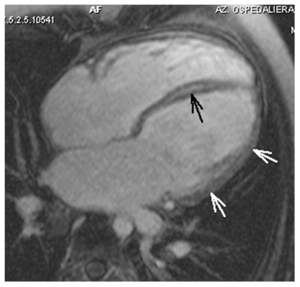
Figure 3. Case 5: four chambers view.
Image acquired 10 min after gadolinium using a routine TI= 190 msec, which nulls normal myocardium at 10 min on our 1T unit. Mild diffuse homogeneous LGE in the LV lateral wall (white arrows), and thick midwall stria in the septum (black arrow). Nulling time at 10 min in this patient, as derived by TI scout sequence, was 113 msec. The data indicate slow gadolinium wash out, suggesting severe diffuse fibrosis.

