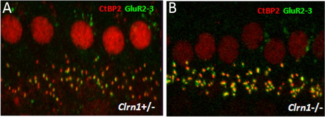
Figure 11.
Light microscopy analysis of ribbon synapse in Clrn1−/− mice. Whole mounts of organs of Corti from P15 mice are double immunolabeled for CtBP2 and GluR2/3 and analyzed by confocal microscopy, followed by deconvolution and surface reconstruction. The difference in the number of double-labeled spots in Clrn1+/− (A) and Clrn1−/− (B) mice was not significant (for quantitative analysis, see Table 2).

