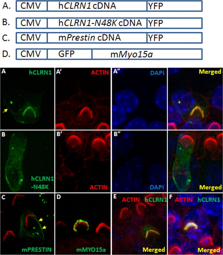
Figure 5.

CLRN1 localizes to the hair bundle. P3 wild-type mouse cochlear epithelia explants transfected with a specific construct (using gene gun) and neighboring nontransfected hair cells after 1 d in culture and ∼20 h after transfection. Expression is driven by the CMV promoter in all cases, and detection of the fluorescent protein marks transfected cells; the tissue was counterstained with phalloidin-conjugated Alexa Fluor 546 (red) and DAPI (blue). A, CLRN1–YFP (green); A′ and A″ are phalloidin and DAPI counterstained images of A. CLRN1–YFP predominantly localized to the bundle with a small amount of the fusion protein localizing to the plasma membrane in the soma (arrow). B, CLRN1N48K–YFP (green); B′ and B″ are phalloidin and DAPI counterstained images of B. C, Merged image showing Prestin–YFP (green)-expressing hair cell and phalloidin-labeled bundles on transfected and nontransfected cells. Arrows point to membrane localization of Prestin–YFP. D, GFP–Myo15a (green)-expressing hair cell and phalloidin-labeled bundles on transfected and nontransfected cells. GFP–Myo15a appears at the tip of the stereocilia as expected. E, F, YFP/phalloidin merged images from two separate (from A) rounds of transfection with CLRN1–YFP construct to show reproducibility of results shown in A.
