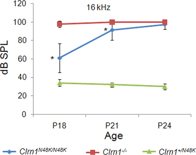
Figure 8.
Assessment of hearing in Clrn1N48/K/N48K knock-in compared with the Clrn1−/− mice over time. The plot shows that mean ABR thresholds of Clrn1N48K/N48K mice (n = 25) are significantly elevated compared with the thresholds of Clrn1+/N48K mice (n = 6), which displayed wild-type thresholds at all time points tested. The plot also shows that ABR thresholds of Clrn1N48K/N48K mice are significantly lower compared with Clrn1−/− mice (n = 12) at P18 and P21; the difference is not significant at P24 because mice from both groups display profound hearing loss at P24. *p < 0.0001.

