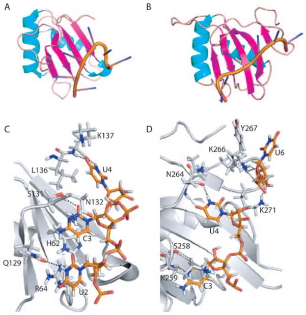
Figure 2.
A and B. Ribbon representations of PTB RRMs 1 and 2 bound to a CUCUCU hexamer. The alpha helices are colored cyan, beta strands magenta and loops beige. C and D. Base specific contacts made by RRMs 1 and 2. Amino acids and nucleotides are shown as stick models. The main chain cartoon traces are colored gray. Atomic contacts are indicated by dashed lines.

