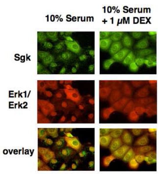
Fig. 1.
Effects of serum and glucocorticoids on the co-localization of Sgk and Erk/MAPK. Con8 mammary tumor cells were treated with 10% serum in the presence or absence of 1μM dexamethasone for 24 hours. The subcellular distribution of Sgk was examined by indirect immunofluorescence microscopy using affinity-purified rabbit polyclonal antibodies to Sgk followed by FITC-conjugated goat anti-rabbit secondary antibodies (green fluorescent staining). Erk1/Erk2 were detected using monoclonal antibodies that specifically recognized both MAPK family members followed by a rhodamine-conjugated anti-mouse secondary antibody employed to selectively visualize this protein kinase (red fluorescent staining). The orange color in the lower panels show the overlap of the green FITC staining and the red rhodamine signal.

