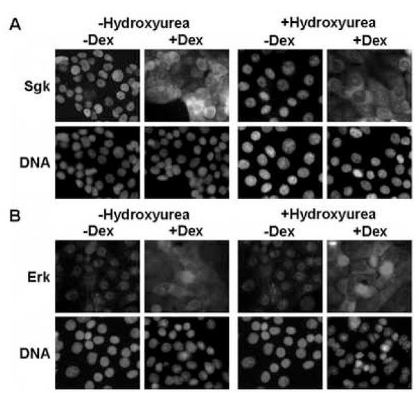
Fig. 2.
Effects of hydroxyurea on the signal-dependent colocalization of Sgk and Erk/MAPK. Con8 cells were treated with 10% serum in the presence or absence of 1 mM hydroxyurea for 24 hours to induce a cell cycle arrest prior to treatment with 1 μM dexamethasone (Dex) for 24 hours. Panel A: The subcellular distribution of Sgk was examined by indirect immunofluorescence microscopy using affinity-purified rabbit polyclonal antibodies to Sgk followed by FITC-conjugated goat anti-rabbit secondary antibodies. Panel B: Erk1/Erk2 were detected using monoclonal antibodies that specifically recognized both MAPK family members followed by a rhodamine-conjugated anti-rabbit secondary antibody employed to selectively visualize this protein kinase. DNA was stained to visualize the nucleus using DAPI.

