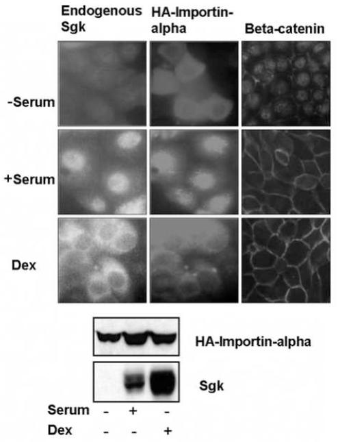
Fig. 3.
Stimulus-dependent localization of importin-alpha and endogenous Sgk. Low confluent (30%) mammary epithelial cells, grown on 2 well lab-tek slides were transfected with expression vectors encoding full length importin-alpha (HA-FLIa) using lipofectamine. The cells were serum starved for 36 hours, and then maintained without serum (−serum) or treated with either 10% calf serum (+ serum) or with 1 μM dexamethasone (Dex) for 15 hours. The localization of HA-importin-alpha (middle panels) and of endogenous Sgk (left panels) and beta-catenin (right panels) was examined by indirect immunoflorescence microscopy as described in the Materials and Methods section. Protein expression of HA-importin-alpha and endogenous Sgk under each of treatment conditions used for localization studies was evaluated by immunoblotting with anti-HA or anti-Sgk antibodies respectively.

