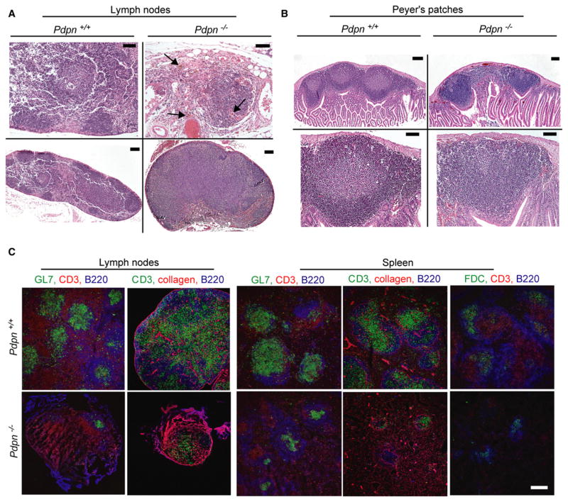
Figure 6. Pdp-Deficient Mice Have a Defect in the Formation of Secondary Lymphoid Structures.
(A) Hematoxylin and eosin staining of paraffin sections of a LN harvested from a Pdpn+/+ mouse (left), a LN remnant harvested from a Pdpn−/− mouse (top right), and a rare normal-sized LN harvested from a Pdpn−/− mouse (bottom right). Lymphocytes stain dark blue and extravasated red blood cells stain red (arrows, top right).
(B) Hematoxylin and eosin staining of a paraffin section of PP harvested from Pdpn+/+ mice (left) and Pdpn−/− mice (right). Top panels show whole PP, and bottom panels show one follicle of the PP.
(C) Cryosections from LNs, LN remnants, and spleens of 12-month-old Pdpn−/− mice and their Pdpn+/+ littermates were analyzed for GC structure via T cell-specific (CD3), B cell-specific (B220), and FDC-specific stains (αCR1, clone 8C12), as well as the GC marker GL7 and the structural marker collagen.
Data shown are representative of three independent experiments. Scale bars in (A) (top) and (B) (bottom) represent 50 μm. Scale bars in (A) (bottom) and (B) (top) represent 100 μm. Scale bar in (C) represents 200 μm.

