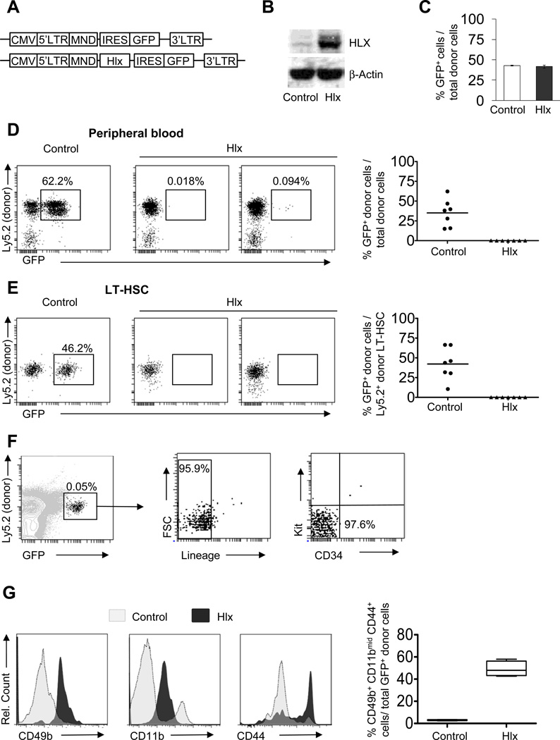
Figure 1. HLX overexpression impairs hematopoietic reconstitution and leads to a decrease in long-term hematopoietic stem cells and persistence of a small progenitor population.
(A) Schematics of lentiviral vectors. (B) Increased protein expression of HLX in Lin−Kit+ cells after transduction with HLX-expressing lentivirus and sorting of GFP+ cells. (C) Homing is not affected by HLX overexpression. 8×10⁴ lentivirus-transduced Lin−Kit+ cells from WT C57BL/6 mice (Ly5.2) were transplanted into lethally irradiated congenic WT recipients (Ly5.1). Bone marrow mononuclear cells from recipients were analyzed 24 hours after transplantation. The frequency of GFP+ cells in the donor population (Ly5.1− Ly5.2+) was assessed and averages +/− SD are shown (N=3). (D, E) Control- or Hlx-IRES-GFP-transduced Lin−Kit+ cells (Ly5.2) together with spleen cells from congenic WT mice (Ly5.1) were transplanted into lethally irradiated congenic WT recipients (Ly5.1) (N=7) and analyzed 12 weeks after transplantation. Total GFP+ cells in peripheral blood (D), and Lin−Kit+Sca+Thy1loFlk2− LT-HSC in bone marrow (E) are shown. Detailed gating scheme and additional analyses are shown in Fig. S1. Means are indicated by horizontal lines in the panels on the right. (F) Analysis of GFP+ cells in total bone marrow cells from recipients transplanted with Hlx-transduced Lin−Kit+ cells after 12 weeks. Relative percentages of GFP+, Lin−, as well as CD34−Kit− donor cells are indicated. (G) CD49b, CD11b, and CD44 expression on donor Ly5.2+GFP+Lin−Kit− cells from recipients transplanted with Hlx- or control-transduced Lin−Kit+ cells 6 weeks after transplantation. Representative histogram plots are shown on the left. Percent of Ly5.2+GFP+Lin−Kit− cells that co-express CD49b+, CD11bmid, and CD44+ are displayed for the control and Hlx groups in the right panel (N=4/condition).

