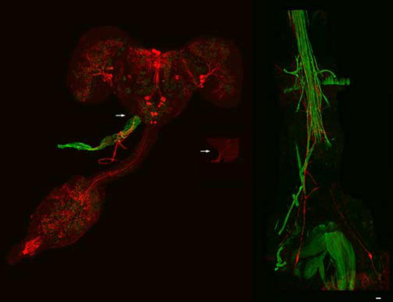
Fig. 1.

Dromyosuppressin (DMS) immunoreactive fibers innervate the aorta and the heart of the blowfly. Neurons (far left; enlargement, shown in center) were stained by fluorescently-labelled DMS antisera in the adult brain (cyanine; red) from which immunoreactive fibers projected to innervate the anterior of the dorsal vessel (arrows). Fluorescently-labelled phalloidin (fluorescein; green) was used to enhance the background in order to view the dorsal vessel. Bilaterally symmetric cells (far right) were stained by DMS antisera from which immunoreactive fibers projected to innervate the posterior of the dorsal vessel. Scale bar = 50 μM.
