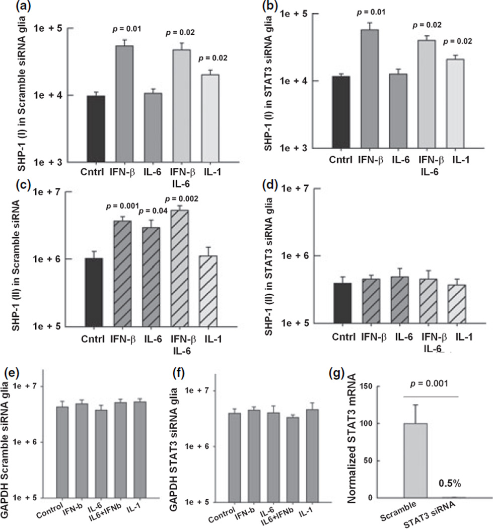
Fig. 8.
SHP-1 promoter I and II expression in STAT-3 siRNA-treated glia. Real-time PCR was used to quantify the expression of the SHP-1 transcripts (mRNA copy number/1.0 ng total RNA) in mixed glia cultures from wild-type mice pre-treated for 72 h with either siRNA agaist STAT-3 or scrambled sequence siRNA. Then, glia were treated for 24 h with media alone, 100 U/mL of IFN-β, 10 ng/mL of IL-6, and 10 ng/mL of IL-1β. (a) SHP-1 (I) in wild-type glial cells pre-treated with scramble siRNA (b). SHP-1 (I) in glia pre-treated with siRNA against STAT-3. (c) SHP-1 (II) in wild-type glia cells pre-treated with scramble siRNA (d) SHP-1 (II) in glia pre-treated with siRNA against STAT-3. (e and f) The levels of GAPDH were quantified in the same samples. (g) The efficiency of the siRNA to deplete STAT-3 mRNA was verified by real time RT-PCR.

