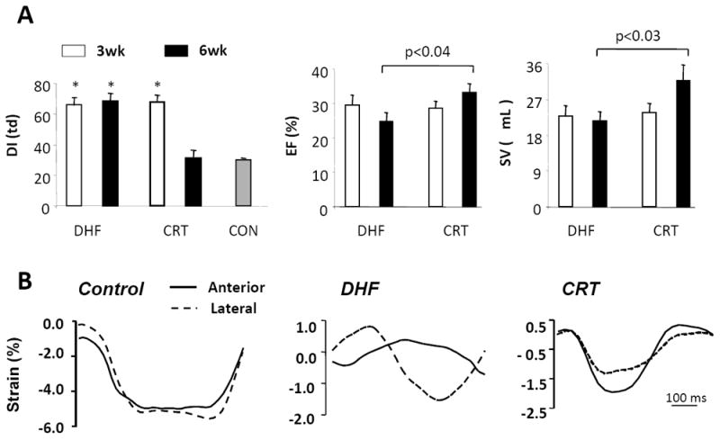
Figure 1.

Development of a canine model of cardiac dyssynchrony and resynchronization. In this model, both dyssynchronous heart failure (DHF) and resynchronized heart failure [cardiac resynchronization therapy (CRT)] groups are exposed first to tachypacing for 3 weeks (in the presence of a preestablished left bundle branch block). This protocol is continued for an additional 3 weeks in DHF hearts, but is switched to rapid biventricular pacing in CRT hearts. A: Left ventricular dyssynchrony (DI, left) is assessed by the variance in timing delay in systolic motion using tissue Doppler. This is similar in both groups at 3 weeks and declines to control levels (synchrony) only in the CRT group. Slight improvements in ejection fraction (EF, middle) and stroke volume (SV, right) are noted in the CRT group, but not in the DHF group at 6 weeks. B: Myocardial strain patterns (tissue Doppler images) obtained in the anterior and lateral wall (tissue Doppler) from a normal control, DHF heart, and CRT heart. With DHF, major disparities in the timing of shortening and reciprocal shortening/stretch in each region are ameliorated by CRT. (adapted from Chakir et al. 22).
