Figure 1.

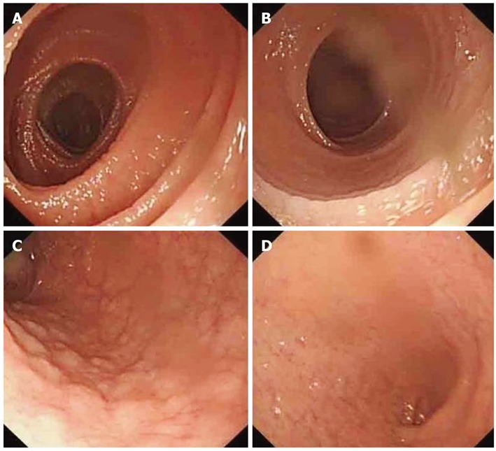
Proximal jejunum mucosa showing scalloping (A), reduced loss of mucosal folds (B), nodularity and mosaic appearance (C) and total mucosal atrophy (D), seen in patients with celiac disease during enteroscopy.

Proximal jejunum mucosa showing scalloping (A), reduced loss of mucosal folds (B), nodularity and mosaic appearance (C) and total mucosal atrophy (D), seen in patients with celiac disease during enteroscopy.